Urinalysis
Sections
Urine Collection & Preparation
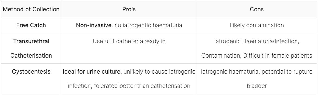
Collection Methods
Preparing Urine for Exam
Dry Mount
1. Centrifuge urine at 1000-1500 rpm for 5 minutes2. Remove fluid above the sediment, leaving a small amount behind3. Remove the sediment and place onto a microscope slide4. Use a second slide to spread the material5. Allow the slide to air-dry6. Use Diff-Quik to stain or send off for external lab prep
Wet Mount
Cells, crystals and casts, will form a sediment due to gravity, mix the whole urine sample well before removing the fluid to be placed in the centrifuge to ensure that the urine sediment wet mount is representative of the whole sample.1. Use a standard volume aliquot of urine for centrifugation (e.g., 5ml). Use a standard volume of urine supernatant to gently resuspend the sediment pellet (i.e. 10% of the original aliquot volume).2. If 5 mL of urine is centrifuged, remove 4.5 mL of supernatant and resuspend the pellet in the remaining 0.5 mL (10%)3. Examine the whole coverslip in 10 fields (areas) per magnification, using both the 10× and 40× objectives4. Use the unstained wet mount to assess concentration of materials within the sediment, because the addition of stain will dilute the sediment.5. Staining is helpful for identification of cell with nuclei but may form crystals or may be contaminated with microorganisms.
6. If crystals or microorganisms are seen confirm they are also there in an unstained wet mount of the sediment
Examining Urine
10 x Magnification
Assess the wet-mount using the 10x objective to find the focus and look for larger structures.
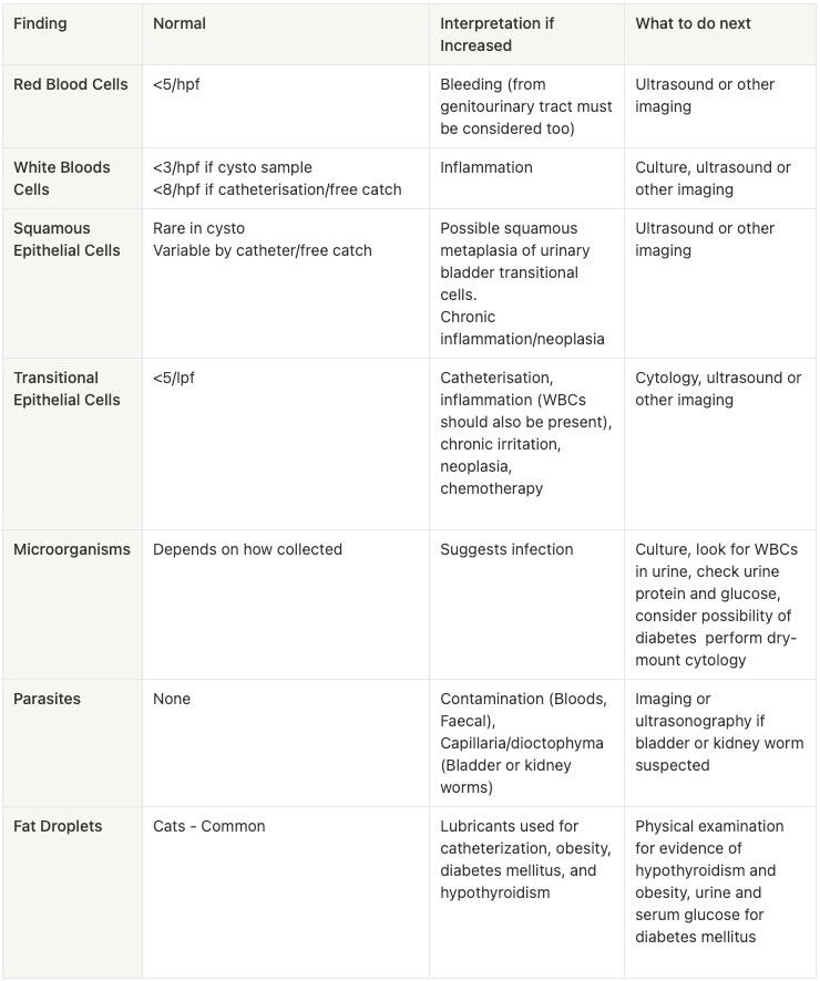
Examine 10 microscopic fields of the slide from low power using the 10x objective for the presence of the following, and provide an assessment of their amounts in the unstained wet mount:Crystals
1. Record type(s) present.
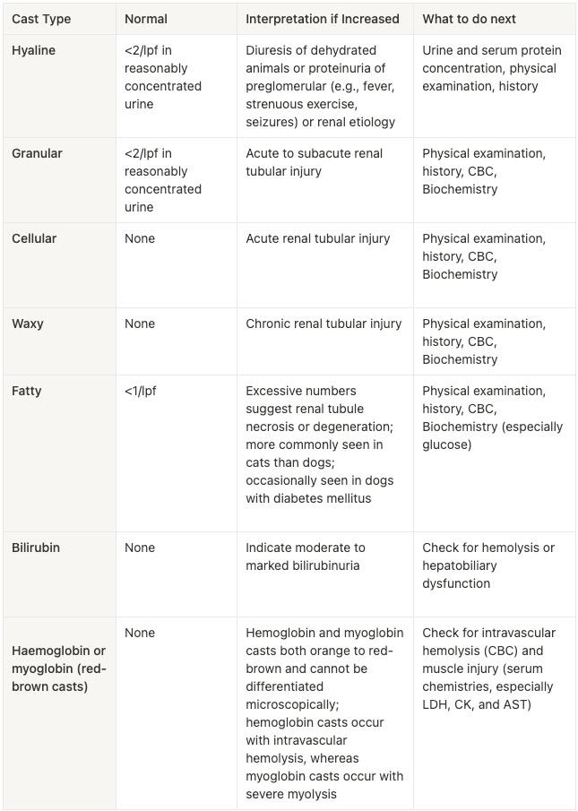
2. Record qualitative impression of the amount of each type of crystal (i.e., none, few, moderate, or many)Casts
1. Normally seen better at the edges of the coverslip.
2. Record type(s) present (e.g., hyaline, cellular, granular, waxy).
3. Record the number of each cast type per 10x low-power field (Ipf) as a range (e.g., 0-2/pt).Epithelial cells
1. Record types present.
2. Record the number of each type of epithelial cell per 10x Ipf as a range (e.g., 0-4/pt).
3. Observe how they look for the presence of dysplastic or neoplastic
changes.Mucous threads
1. Record qualitative impression of the number of mucous threads (none, few, moderate, or many).Worm eggs, larva, adult worms, or other parasites
1. Record qualitative impression of the number of parasite structures
(none, few, moderate, or many).
40x Magnification
Examine 10 fields - assess the morphology of structures observed at 10x magnificationConfirm the identification of the structures observed at low power. Inspect epithelial cells for dysplastic or neoplastic cytomorphology Inspect the slide for the presence of the following and assess the number of each in the unstained wet mount:Red Blood Cells (Erythrocytes):1. Record the number of redblood cells per high-power field (hpf) as a range (e.g., 2-4/hpf)Leukocytes (White Blood Cells):1. Record the number of leukocytes as a range (e.g., 0-3/hpt)Microorganisms (e.g., bacteria, yeast, fungi, algae)1. Record morphology of bacteria (e.g.. cocci, bacilli, filamentous, spore-forming).
Record qualitative impression of the number of each type of microorganism (none, few, moderate, or many)Lipid droplets
1. Lipid droplets need to be distinguished from erythrocytes
2. Lipid droplets are variably sized, are refractive, and often float above the focal plane.
3. Record qualitative impression of the number of lipid droplets (none, few, moderate, or many).Other structures or unidentified structures1. Identify the structures or describe their morphology
2. Record qualitative impression of the number of these structures (none, few, moderate, or many).
Interpretation of Findings